Glaucoma
The Difference Between Retinal Conditions and Glaucoma

Mr Minak Bhalla
From recognising familiar faces to appreciating the beauty of the natural world, our experience of life is shaped profoundly by vision. Eye conditions can affect different structures within the eye — including the retina, which captures light and converts it into signals, and the optic nerve, which carries those signals to the brain. Damage to these structures can give rise to different eye diseases, including glaucoma and retinal conditions, which can sometimes produce overlapping symptoms. Understanding the differences between them, and seeking early diagnosis, is essential for protecting your sight.
Understanding Retinal Conditions
Retinal conditions affect the retina — the light-sensitive inner layer at the back of the eye. Some common retinal conditions include:
- Retinal detachment
- Diabetic retinopathy
- Age-related macular degeneration
- Retinal tears
- Retinal vascular disorders
Symptoms of retinal conditions may include:
- Sudden flashes of light
- Floaters (small spots or shadows drifting across vision)
- A curtain-like shadow across part of the visual field
- Distorted vision, or objects appearing smaller or larger than expected
- Blurred vision
- Sudden or progressive vision loss
Understanding Glaucoma
Glaucoma is a group of eye conditions characterised by progressive damage to the optic nerve, most commonly associated with raised intraocular pressure (IOP). The optic nerve damage caused by glaucoma is irreversible. The most common form — chronic open-angle glaucoma — tends to develop gradually and silently, affecting peripheral vision first.
Symptoms may include:
- Gradual loss of peripheral (side) vision
- Blurred vision in advanced disease
- In acute angle-closure glaucoma: sudden severe eye pain, headache, halos around lights, nausea, and vomiting — this is an ocular emergency requiring immediate attention
What are the Differences? Retinal Conditions vs Glaucoma
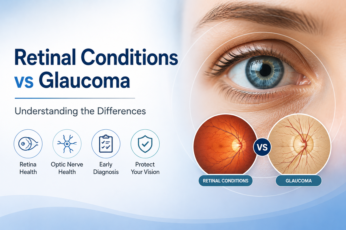
Although both glaucoma and retinal conditions can cause visual disturbance, the fundamental distinction is the structure of the eye that is affected.
Glaucoma
- Affects the optic nerve
- Usually associated with raised pressure inside the eye
- Typically causes a slow, often unnoticed loss of peripheral vision
- Often called the "silent thief of sight" because significant damage can occur before symptoms are noticed
Retinal conditions
- Affect the retina
- May develop due to ageing, diabetes, injury, or other eye disorders
- Often cause more sudden symptoms such as flashes, floaters, shadows, or loss of vision
A thorough eye examination by an ophthalmologist can distinguish between the two and guide appropriate treatment.
Can Retinal Disease be Confused with Glaucoma?
Yes. Both conditions can affect vision, and some symptoms overlap. Shared features may include:
- Blurred vision or difficulty seeing clearly
- Difficulty with daily activities due to poor vision
- Gradual changes in vision
- Difficulty seeing in low light
- Disturbances in the visual field
However, the underlying causes are distinct. In glaucoma, vision loss results from progressive optic nerve damage, whereas in retinal disease it results from changes to the retina itself.
Ophthalmologists use several investigations to identify the cause of visual symptoms, including:
- Measurement of intraocular pressure
- Optic nerve examination
- Retinal examination
- Visual field testing
- Imaging of the eye (OCT and fundus photography)
Do Glaucoma and Retinal Disease Present with Similar Symptoms?
There is some overlap, but important differences exist. Glaucoma — particularly the most common open-angle form — typically causes slow, progressive peripheral vision loss that patients often do not notice until the disease is advanced. Retinal conditions more commonly produce sudden symptoms such as flashes of light, new floaters, or dark shadows in the visual field.
Awareness of these distinctions supports earlier presentation and diagnosis.
When Should You See a Retinal Specialist?
A retinal specialist is an ophthalmologist with expertise in diagnosing and treating conditions affecting the retina and the back of the eye.
You should seek a prompt retinal assessment if you experience:
- Sudden vision loss
- New flashes of light
- A sudden increase in floaters
- A dark shadow or curtain in the visual field
- Distorted vision
- Changes in vision in the context of diabetes
- A history of eye or retinal injury
Early assessment of retinal symptoms can help preserve remaining vision and reduce the risk of complications.
Management of Retinal Conditions
Monitoring and Eye Examinations
Some retinal conditions do not require immediate treatment but benefit from regular monitoring through eye examinations, retinal imaging, and vision testing to determine whether intervention is needed.
Medical Management
Some retinal conditions can be managed with medication. For example:
- Anti-VEGF (vascular endothelial growth factor) injections are used in the treatment of wet age-related macular degeneration and diabetic macular oedema, where they reduce abnormal blood vessel growth and leakage
- Steroid medications may be used in inflammatory retinal conditions
Laser Treatment
Laser therapy may be recommended for conditions such as diabetic retinopathy and retinal tears. It can seal abnormal blood vessels and reduce the risk of retinal detachment.
Surgery
Structural retinal problems may require surgical management:
- Retinal detachment: vitrectomy, scleral buckling, or pneumatic retinopexy to reattach the retina
- Retinal tears: laser treatment or cryotherapy (freezing treatment) to seal the tear
When Should You See a Glaucoma Specialist?
A glaucoma specialist is an ophthalmologist with expertise in managing conditions affecting the optic nerve and intraocular pressure.
You should consider consulting a glaucoma specialist if you have:
- Raised intraocular pressure
- A family history of glaucoma
- Changes in peripheral vision
- Optic nerve abnormalities detected on eye examination
- A history of eye injury or prior eye surgery
- Difficulty adapting to low light
As glaucoma can cause permanent vision loss before producing noticeable symptoms, regular eye screening is strongly recommended — particularly for those with risk factors.
Management of Glaucoma
Treatment aims to reduce intraocular pressure and prevent further damage to the optic nerve.
1. Medical Management (Eye Drops)
Eye drops are a mainstay of glaucoma treatment and may be recommended as a first-line option or alongside laser treatment. Prostaglandin analogue (PGA) eye drops are the recommended first-line topical treatment, with beta-blockers, carbonic anhydrase inhibitors, and alpha-agonists used as second and third-line options or in combination. Consistent and correct use of prescribed drops is important for effective pressure control.
2. Laser Treatment
Selective laser trabeculoplasty (SLT) is now recommended by NICE as a first-line treatment option for open-angle glaucoma and ocular hypertension. It uses targeted laser energy to improve the drainage of fluid from the eye through the trabecular meshwork, thereby reducing intraocular pressure. It is a minimally invasive outpatient procedure. Other laser options include laser iridotomy (for angle-closure glaucoma) and laser cycloablation.
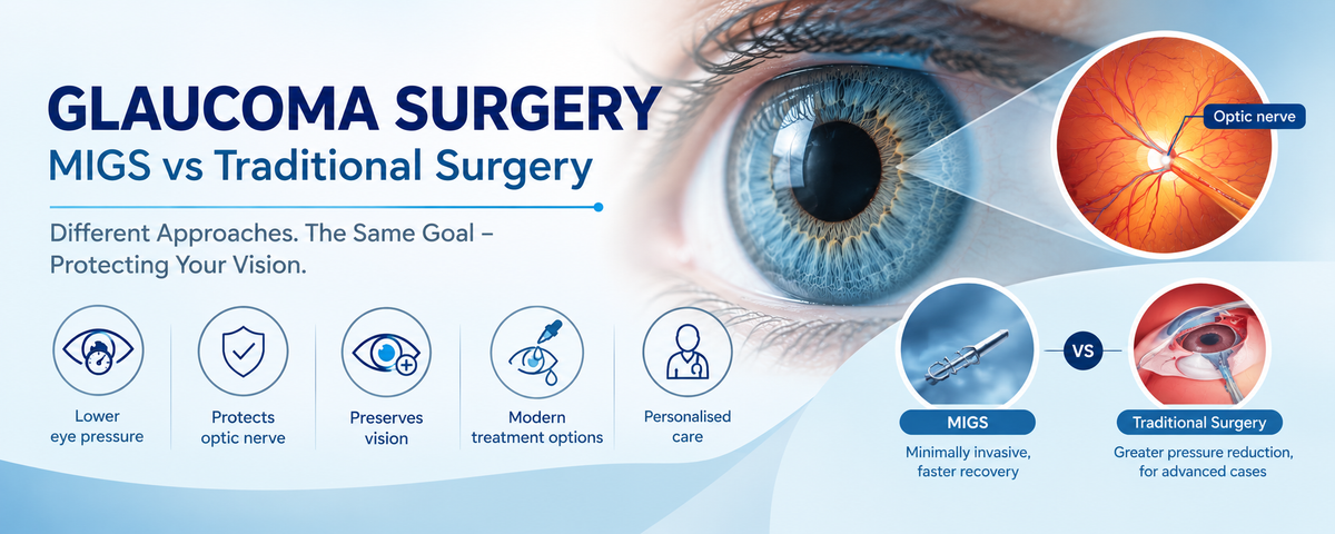
3. Minimally Invasive Glaucoma Surgery (MIGS)
MIGS encompasses a group of surgical procedures performed through small incisions using microscopic devices to improve aqueous outflow from the eye. It offers a lower-risk surgical option compared to traditional glaucoma surgery. Widely used MIGS devices include the iStent, Hydrus microstent, and the OMNI surgical system.
4. Trabeculectomy
Trabeculectomy is the most established surgical option for advanced glaucoma. A small opening is created in the trabecular meshwork to form a new drainage pathway for aqueous humour, reducing intraocular pressure.
5. Preserflo Microshunt
The Preserflo microshunt is an advanced minimally invasive device that drains aqueous humour from the anterior chamber to the subconjunctival space, reducing intraocular pressure with a less invasive approach than traditional filtration surgery.
6. Aqueous Shunt Surgery
Aqueous shunt surgery is typically considered when intraocular pressure remains uncontrolled despite medical and laser treatment, or following a failed trabeculectomy. A small drainage implant is placed in the eye to divert aqueous humour to an external reservoir, lowering intraocular pressure in moderate to severe cases.
Why are Routine Eye Examinations Important?
Many eye conditions produce no symptoms until significant damage has already occurred. This is particularly true of glaucoma. Regular eye examinations enable early detection of changes in the optic nerve, retina, and intraocular pressure — allowing treatment to begin before vision is affected.
Conclusion
If you are experiencing changes in your vision and would like to understand whether they may be related to glaucoma or a retinal condition, an assessment by an experienced ophthalmologist is the most important step. Mr. Minak Bhalla is a consultant ophthalmologist and glaucoma specialist based at Harley Street, London, with over ten years of experience in advanced glaucoma surgery. He provides personalised eye assessments and specialist care for glaucoma and related eye conditions. Book your appointment at The London Clinic, 119 Harley Street, London W1G 6AU, or at Clinica London, 1st and 2nd Floor, 140 Harley Street, London W1G 7LB.
Frequently Asked Questions - Retinal Conditions vs Glaucoma
1. What is the main difference between glaucoma and retinal disease?*
Glaucoma primarily affects the optic nerve, typically due to raised pressure inside the eye, while retinal diseases affect the retina — the light-sensitive layer at the back of the eye. Both can cause vision problems but involve different structures and require different diagnostic and treatment approaches.
2. Can retinal disease be mistaken for glaucoma?
Yes. Both may cause blurred vision, difficulty performing daily activities, and changes in the visual field. A detailed eye examination — including optic nerve assessment, retinal imaging, intraocular pressure measurement, and visual field testing — helps determine whether symptoms are related to the retina or the optic nerve.
3. What are the warning signs of retinal problems?
Common warning signs include sudden flashes of light, new floaters, a curtain-like shadow across part of the visual field, distorted vision, and sudden changes in eyesight. These symptoms warrant prompt assessment by an ophthalmologist.
4. Can I consult a glaucoma specialist at Harley Street, London?
Mr. Minak Bhalla is an experienced consultant ophthalmologist and glaucoma specialist at Harley Street, London, with over ten years of expertise in advanced glaucoma procedures including MIGS, trabeculectomy, Preserflo microshunt, and aqueous shunt surgery. Book a consultation today for a thorough assessment and personalised treatment plan.