Glaucoma
Glaucoma: What It Is, Symptoms, Diagnosis and Treatment

Dr Minak Bhalla
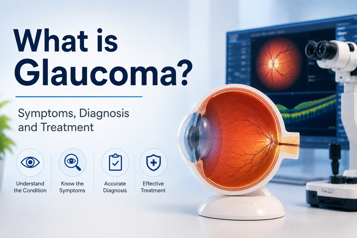
What is Glaucoma?
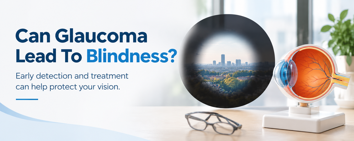
Glaucoma is a group of eye conditions characterised by progressive damage to the optic nerve. This damage is most commonly associated with raised intraocular pressure (IOP), although glaucoma can occur even when pressure is within the normal range. Critically, optic nerve damage caused by glaucoma is irreversible, and vision loss cannot be recovered once it has occurred. This makes early diagnosis and prompt management essential.
In the most common form — primary open-angle glaucoma — the condition is largely asymptomatic in its early stages. Around half of all people living with glaucoma are unaware they have the condition, as significant nerve damage can occur before any symptoms are noticed. This is why glaucoma is often referred to as the "silent thief of sight," and why regular eye examinations are so important, particularly for those at higher risk.
Types of Glaucoma
Congenital or Developmental Glaucoma
- Primary congenital glaucoma: caused by a primary developmental abnormality in the drainage angle of the eye
- Secondary congenital glaucoma: associated with other ocular or systemic disorders
Acquired Glaucoma
Primary acquired glaucoma:
- Primary open-angle glaucoma (POAG): the most common type. The drainage angle remains open, but partial blockage of the trabecular meshwork gradually impairs aqueous outflow, increasing pressure and causing progressive optic nerve damage. It typically develops slowly and without symptoms until advanced disease
- Primary angle-closure glaucoma: the drainage angle becomes narrowed or anatomically closed. Intraocular pressure can rise rapidly, particularly when the pupil dilates, as the peripheral iris obstructs the drainage angle. Acute angle-closure is an ocular emergency
- Secondary acquired glaucoma: caused by other ocular conditions such as eye injury, trauma, iritis, retinal detachment, or cataract, or by systemic conditions including diabetes.
Who is at Risk of Glaucoma?
Risk factors for glaucoma include:
- Age over 40, with risk increasing with each decade of life
- Family history of glaucoma
- Diabetes
- Reduced corneal thickness
- Short-sightedness (myopia)
- A history of eye injury or previous eye surgery
- Early oestrogen deficiency
- Long-term use of corticosteroid medication
Symptoms of Glaucoma
Open-angle glaucoma:
Open-angle glaucoma is often asymptomatic in the early stages. As the condition progresses, symptoms may include:
- Patchy blind spots in the visual field
- Loss of peripheral (side) vision
- Blurred vision in advanced disease
Angle-closure glaucoma (acute):
Acute angle-closure glaucoma is an emergency. Symptoms include:
- Severe eye pain
- Severe headache
- Nausea and vomiting
- Sudden blurred vision
- Halos around lights
- Eye redness
If you experience these symptoms, seek urgent medical attention.
Diagnosing Glaucoma
Several investigations are used to diagnose and monitor glaucoma:
Tonometry
Tonometry measures intraocular pressure. It is performed either by directing a small puff of air at the eye or by gently touching the front of the eye with an instrument after the application of anaesthetic eye drops. It is quick and painless.
Pachymetry
Pachymetry measures corneal thickness using an ultrasound probe or optical scanner. Corneal thickness can influence IOP readings and risk assessment.
Gonioscopy
Gonioscopy examines the drainage angle of the eye — the area where aqueous humour leaves the anterior chamber. After anaesthetic eye drops are applied, a special mirrored lens is placed on the eye, enabling the ophthalmologist to determine whether the drainage angle is open, narrow, or closed.
Ophthalmoscopy
Ophthalmoscopy examines the retina and optic nerve at the back of the eye using a bright light and magnifying instrument. It allows assessment of the optic disc for signs of glaucomatous damage.
Visual Field Testing (Perimetry)
This test assesses peripheral vision by asking the patient to focus on a central target and indicate when they detect lights appearing in different areas of the visual field.
Optical Coherence Tomography (OCT)
OCT uses light waves to produce cross-sectional images of the retina and optic nerve. It can detect thinning of the retinal nerve fibre layer and other structural changes that may indicate glaucoma before symptoms develop.
Treatment of Glaucoma
All treatment is directed at reducing intraocular pressure to prevent further optic nerve damage. The most appropriate approach depends on the type and severity of glaucoma, the patient's individual circumstances, and their response to treatment.
1. Medical Management (Eye Drops)
Eye drops are a cornerstone of glaucoma treatment. Prostaglandin analogue (PGA) eye drops — such as latanoprost — are the recommended first-line topical treatment, as they are the most effective class for lowering IOP and require only once-daily dosing. Beta-blockers, carbonic anhydrase inhibitors, and alpha-agonists are used as second and third-line agents, either in addition to or as alternatives to PGAs when these are not tolerated. Consistent use of prescribed drops is essential, as their benefit is preventative rather than immediately noticeable to the patient.
2. Laser Treatment
Selective laser trabeculoplasty (SLT) is a minimally invasive outpatient procedure that uses targeted laser energy to improve aqueous drainage through the trabecular meshwork. It is now recommended as a first-line treatment option for open-angle glaucoma and ocular hypertension, either as an alternative to or alongside eye drops. Other laser options include laser iridotomy for angle-closure glaucoma and laser cycloablation.
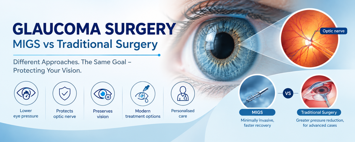
3. Minimally Invasive Glaucoma Surgery (MIGS)
MIGS encompasses a group of procedures performed through small incisions using microscopic devices to improve aqueous outflow. It offers a lower-risk surgical option compared to traditional glaucoma surgery. Widely used devices include:
- iStent: a small titanium stent implanted into the trabecular meshwork to improve aqueous outflow
- Hydrus microstent: a flexible microstent placed to dilate Schlemm's canal and enhance aqueous drainage
- OMNI surgical system: combines canal dilation and trabeculotomy to enhance drainage through the eye's natural outflow pathway
4. Trabeculectomy
Trabeculectomy is the most established surgical option for advanced glaucoma not adequately controlled by drops or less invasive procedures. A small opening is created in the trabecular meshwork, forming a new drainage pathway for aqueous humour. A small collecting reservoir called a filtering bleb is created from conjunctival tissue, allowing fluid to leave the eye and be absorbed into the circulation, thereby reducing IOP.
5. Preserflo Microshunt
The Preserflo microshunt is an advanced minimally invasive implant that drains aqueous humour from the anterior chamber to the subconjunctival space, lowering IOP with a less invasive approach than traditional filtration surgery.
6. Aqueous Shunt Surgery
Aqueous shunt surgery is typically recommended when IOP remains uncontrolled despite medical and laser treatment, or following a failed trabeculectomy. A drainage implant is placed in the eye to divert aqueous humour to an external reservoir, reducing pressure in moderate to severe cases.
Consultation with an experienced glaucoma surgeon is important in determining the most appropriate management approach for each individual patient.
Looking for a Glaucoma Surgeon at Harley Street?
If you are experiencing symptoms such as blurred vision, eye pain, or halos around lights, or if you have been told you are at risk of glaucoma, booking a prompt assessment with an experienced ophthalmologist is an important first step. Mr. Minak Bhalla is a consultant ophthalmologist and glaucoma surgeon at Harley Street, London, with over ten years of experience and more than 2,000 glaucoma and cataract procedures. He offers the latest generation MIGS devices and advanced glaucoma surgery, alongside personalised assessment and treatment tailored to your individual needs. You can reach Mr. Bhalla at The London Clinic, 119 Harley Street, London W1G 6AU, or at Clementine Churchill Hospital, Harrow, HA1 3RX.