Retina
Retinal Vein Occlusion: Causes, Symptoms & Treatment Guide

Dr Minak Bhalla
Vision changes can be sudden, turning regular activities like reading, driving or recognising familiar faces into a challenge. One of the common vision changes that can affect your quality of life is blurring of vision. In some cases, blurred vision can be a sign of retinal vein occlusion. Our eyes rely on a network of several blood vessels to deliver oxygen and nutrients necessary for clear vision. When one of these retinal veins becomes blocked, it can lead to vision loss and other complications- the condition known as retinal vein occlusion.
What is retinal vein occlusion?
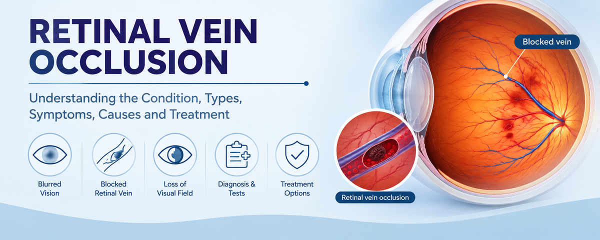
The retina is a thin and light-sensitive layer of tissue at the back of the eye that converts light rays into signals and sends it to the brain, allowing us to see. The retina receives nutrition from retinal arteries, while retinal veins carry blood away from the retina. When a retinal vein becomes blocked due to a blood clot or compression, blood and fluid accumulation in the retina can cause swelling and bleeding within the retina causing impairment of vision.
Retinal vein occlusion may involve the central retinal vein which causes immediate and severe loss of vision, or a branch retinal vein which results in partial loss of vision, depending on the affected region drained by the vein.
Types of retinal vein occlusion
Retinal vein occlusion can be classified into two types:
Central retinal vein occlusion
When the central retinal vein becomes blocked, it is called the central retinal vein occlusion (CRVO). This vein drains blood from the entire retina, so blockage in it can lead to severe vision loss. CRVO can be further classified into -
Non-Ischemic CRVO: It is the milder form of the disease with partial blockage. Although the central retinal vein is blocked, enough blood continues to reach the retina. This usually causes blurred vision but the outcome is better than ischemic CRVO. This accounts for 75% of cases.
Ischemic CRVO: It is the more severe form of CRVO, where the blockage significantly reduces draining of blood out of the retina, eventually resulting in reduced blood and oxygen supply, leading to retinal damage. This accounts for the remaining 25% of cases.
Branch retinal vein occlusion Branch retinal vein occlusion affects one of the smaller branches of the retinal vein. It can occur near the optic disc and affect a major part of the retina, or the part where a retinal vein crosses a retinal artery.
What causes retinal vein occlusion?
Retinal vein occlusion may occur due to the following reasons-
Formation of a blood clot within the retinal vein
Compression of a retinal vein by a retinal artery, at the site where it crosses path with the retinal artery
Increased pressure within the eye
Slowing down of blood flow
Who is more at risk?
Age over 40 years (More common around 60s)
High blood pressure
Diabetes
Increased pressure within the eye due to glaucoma ocular hypertension
Cardiovascular diseases (especially atherosclerosis)
Signs and symptoms of retinal vein occlusion
Non-Ischemic central vein occlusion
Slightly or moderately blurred vision
Gradually or suddenly reduced vision
Distorted vision: Objects may look out of shape or straight lines may appear wavy
Floaters
Ischemic central vein occlusion
Sudden, severe, painless loss of vision
Marked reduction in vision causing difficulty in reading, driving or recognizing faces
Visual field loss
Dense floaters
Difficulty distinguishing objects from their backgrounds, especially in dim light
In advanced cases, patients may later experience:
i. Eye pain
ii. Redness
iii. Headache
iv. Halos around light
Branch retinal vein occlusion
Sudden painless blurred vision
Partial loss of vision or a blind spot in one area of vision field
Distorted vision: Objects may look out of shape or straight lines may appear wavy
Some patients may show no symptoms, in such cases the condition can only be detected during a routine eye examination
How can retinal vein occlusion be diagnosed?
If you suddenly experience any of the symptoms of retinal vein occlusion, it is important to consult an ophthalmologist as soon as possible. An eye examination by an experienced ophthalmologist is essential for diagnosis of the condition.
The following tests are used to diagnose retinal vein occlusion
Comprehensive eye examination
The ophthalmologist may ask about your symptoms and medical history, including any existing condition such as diabetes, high blood pressure, glaucoma or high cholesterol. A vision test is conducted using a standard eye chart to determine how much the vision has been affected.
Dilated eye examination
Eye drops are used to dilate the pupils, allowing the ophthalmologist to get a better view of the retina. Using a specialised microscope, the ophthalmologist looks for signs of retinal vein occlusion, such as:
i. Bleeding within the retina
ii. Swollen retinal veins
iii. Fluid buildup
iv. Spots looking like cotton wool (small areas of retinal damage)
Optical coherence tomography
It is a quick, painless imaging scan showing detailed cross-sectional images of the retina. It helps the ophthalmologist to detect swelling, measure thickness of the retina and assess the extent of damage within the retina.
Fundus fluorescein angiography
In this test, a harmless fluorescent dye is injected into a vein in the arm. As the dye travels through the retinal blood vessels, a specialised camera is used to take pictures showing how the blood is flowing through the retina. It helps to identify blocked blood vessels, leaking blood vessels and areas with poor blood supply.
Additional health assessment
As retinal vein occlusion is often associated with other medical conditions, following health assessments can be recommended -
i. Blood pressure measurement
ii. Blood glucose testing
iii. Cholesterol testing
iv. Blood tests to detect blood clotting disorders
Management of retinal vein occlusion
The treatment depends on the type and severity of retinal vein occlusion, and the presence of complications like macular edema (fluid buildup and swelling in the retina). While the existing damage may not always be reversed, treatment can help improve the vision or prevent it from worsening, reduce complications that can harm your eyes and improve overall eye health.
Anti-VEGF eye injections
It is one of the most common treatments for retinal vein occlusion. VEGF(Vascular endothelial growth factor) is a protein that causes macular edema and abnormal growth of blood vessels. Anti-VEGF injections can prevent that. In this procedure, the eye is numbed with anaesthetic drops before the injection is administered into the eye. This procedure is quick and takes only a few minutes. Some common injections used are Ranibizumab, Bevacizumab and Aflibercept. It can help to-
i. Reduce swelling in the retina
ii. Stop leaking blood vessels
iii. Prevent growth of abnormal blood vessels
iv. Improve or preserve vision
Steroid injections or implants
If retinal swelling does not improve with Anti-VEGF injections or they are not suitable for a patient, steroid treatment can be used. Steroids help to reduce inflammation and swelling in the retina. Depending on your condition, your ophthalmologist will decide whether you need the steroid in the form of an injection or a tiny implant placed in the eye which slowly releases the medicine over time.
Laser treatment or Panretinal photocoagulation (PRP)
In case of abnormal growth of new blood vessels in the eye due to poor blood supply, laser treatment is recommended. A specialised laser is used to create small burns in areas of the retina that lack blood flow. This helps to decrease the number of VEGF proteins which promote the growth of abnormal blood vessels. This laser can -
i. Prevent further bleeding
ii. Reduce risk of serious complications like neurovascular edema (Abnormal
fluid buildup in the brain)
iii. Protect the remaining healthy part of the retina
Vitrectomy surgery
Posterior pars plana vitrectomy is a surgery that may be needed in some cases of retinal vein occlusion, where complications like non-clearing vitreous haemorrhage or fractional retinal detachment develop. It can help patients who experience severe bleeding in the eye that lasts more than four weeks or keeps coming back. In this procedure, the blood filled vitreous gel is removed from the eye and replaced with a clear solution to help the eye heal.
Managing associated health conditions
Along with the treatments for the retinal vein occlusion, it is also recommended to manage the underlying medical conditions that can increase the risk of it. The doctor may advise you to -
i. Keep blood pressure under control
ii. Manage diabetes
iii. Stop smoking
iv. Lower high cholesterol
v. Eat healthy diet and increase physical activity
Conclusion
Retinal vein occlusion is a serious eye condition that needs urgent medical attention. A timely diagnosis and treatment can help to prevent complications and preserve your vision. If you are experiencing symptoms of retinal vein occlusion and looking for an experienced ophthalmologist in Harley Street, we are here to provide the treatment and support you need. Dr. Minak Bhalla is an experienced consultant ophthalmologist in Harley Street with 10 years of experience in treatment of glaucoma, cataract surgery and general ophthalmology. Dr. Bhalla completed his ophthalmological training at Moorfields Eye Hospital, Royal Free Hospital, and Western Eye Hospital, where he developed exquisite skills in complex advanced surgical procedures. He will provide you with personalised retinal examination and assist his colleagues with the administration of injections aiming to treat retinal vein occlusion at Harley Street, London. Reach us at The London Clinic in 119 Harley Street, London W1G 6AU and The Clementine Churchill Hospital in Sudbury Hill, Harrow HA1 3RX to gift your eyes the care that they deserve.